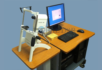
The Heidelberg HRT II Tomographer uses scanning laser tomography to produce the 3-dimensional images, which allow precise measurements of optic nerve head parameters. This technology is purported by Heidelberg Engineering to differentiate between normal eyes and glaucomatous eyes even before visual field defects are present. The Heidelberg HRT II features automatic image acquisition, sector analysis for early glaucoma detection, and automatic progression analysis. Also, the Heidelberg HRT II produces results which are presented in a format that is understood by patients to ensure their compliance. The Heidelberg Retina Tomography operation software provides a variety of functions to quantitatively describe the three-dimensional properties of the retina and the optic nerve head and to precisely detect and describe topographic changes.
The Heidelberg HRT II has a 15° × 15° field of view, a 12 to +12 diopters (automatic) focus range, digitized image size, 2D images of 384×384 pixels, 3D images of 384×384×16 to 384×384×64 pixels, optical resolution transverse 10 µm, longitudinal 300 µm, digital resolution transverse 10 µm/pixels, longitudinal 62 µm/pixels, scan time for 2D images 0.024 seconds, and scan time for 3D images is 0.4 to 1.5 seconds.


