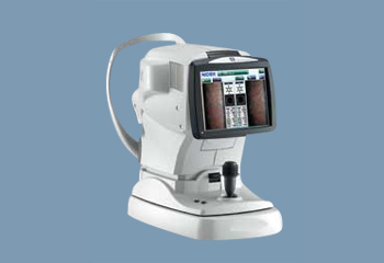
Paracentral Specular Microscopy
In addition to conventional central and peripheral specular microscopy, the CEM-530 includes a unique function to capture paracentral images. The paracentral images are captured at eight points, 5º visual angle within a 0.25 mm x 0.55 mm field and enable enhanced assessment surrounding the central image.
Auto Indication of the Optimal Image
16 images are captured and automatically sorted based on quality and the ability to be analyzed. The optimal image for analysis is indicated with orange highlight.
This feature aids the clinician in the selection of the most suitable image for analysis and enhances reliability of the data.
Two-second Auto Analysis
Rapid analysis increases the efficiency of the practice. Once the image is selected, complete analysis is automatically performed in two seconds with the CEM-530.
The analysis screen allows visualization of the endothelial cells in four modes, trace, photo, area, and apex, which helps the clinician to verify analysis values with the correspondent cell images.


