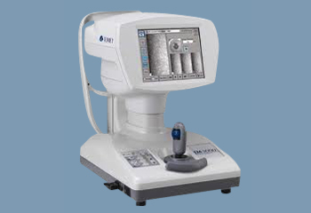
The Specular Microscope EM-3000 provides corneal endothelium imaging by using the specular optical principle with a visible light source, lenses and a CCD camera. These project a slit of light to the cornea from an oblique direction and capture the reflected image of the corneal endothelium by CCD camera. The EM-3000 newly incorporates an LED light source in place of a halogen lamp and a high-speed CCD camera to capture 15 images in series. It automatically selects and instantly displays the finest image of these images. In addition, a built-in clinical application delineates the endothelial cells automatically and displays statistical data such as cell numbers, average area and cell density on the color LCD.


