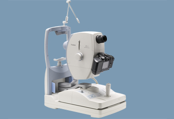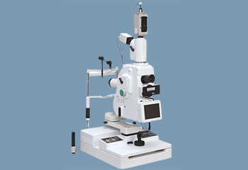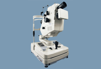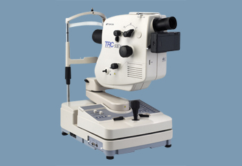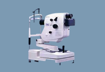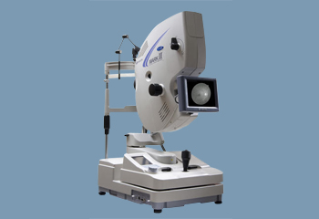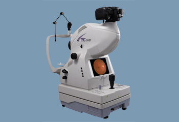
Fundus photography involves the use of a specialized low power microscope that’s attached to a camera. The camera-microscope combination then directly photographs the retina, using the pupil as an entrance and exit point for imaging light rays.
How mydriatic fundus cameras work
Like many pieces of ophthalmology equipment, patients sit at a mydriatic fundus camera and rest their chins on a chinrest and their foreheads against a bar. While they rest in this position, the assigned photographer aligns the fundus camera to capture the best images. Those images are used to diagnose eye diseases as well as treat them and follow their progression. Some common diseases identified and monitored using fundus photographs include glaucoma and diabetic retinopathy. Fundus cameras can also catch retinal issues that are easily missed using fluorescein angiograms.
Benefits of mydriatic fundus cameras
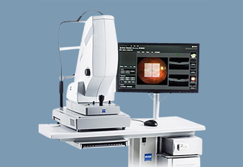
Mydriatic fundus cameras use the retina’s reflective properties to illuminate issues that aren’t detected with common technology like slit lamps.
Because of the specificity of fundus photography, many conditions can be detected early and treated in a way that protects the patient’s long-term eye health.
Also, mydriatic fundus cameras can give the ophthalmologist a more comprehensive view of a patient’s eye health; the detail provided in these photos creates a fuller picture than what can be obtained by patient complaints and other technology or assessment procedures.
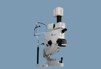
Mydriatic fundus cameras are also remarkably easy to use.
To improve the accuracy of your diagnoses and treatment plans, a mydriatic fundus camera is a worthy addition to your practice.