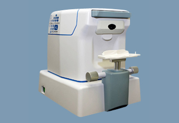
The Konan CellChek XL Specular Microscope captures consistent, high quality images of the patient’s corneal endothelium using a patented method that identifies the position of the cellular interface. The Konan CellChek XL automatically performs final alignment and fine focus, and then captures the image. A copy of the file is auto-archived on an 80GB external hard drive for an additional level of assurance. Access to the Konan CellChek XL computer is controlled through the use of a security key.
The Konan CellChek XL offers five fixation points for image capture at the center and four peripheral sites, allowing a more comprehensive look at the cornea. This is particularly valuable in cases such as DSAEK, ALK, or in the presence of corneal dystrophies.
The software on the Konan CellChek XL allows you to record the location on the cornea where images are captured, review patient history, track changes to the patients’ corneas with time, and link the patient scans to an electronic health record. It also measures corneal thickness at all five target sites using non-contact technology.
Features:
• The Konan CellChek XL is fully automated, which reduces exam and analysis time to less than 30 seconds
• Semi-automated, which allows the user to make subjective enhancements to the automatically-generated cell pattern overlay,
• Center method, which is the standard method used by reading centers, and
• Flex-center method, which is particularly useful for analyzing images containing a small number of cells